There are several theories, including inadequate intake of fibre, prolonged sitting on the toilet, and chronic straining during a bowel movement. None of these theories has strong experimental support. Pregnancy is a clear cause of enlarged haemorrhoids though, again, the reason is not clear.
As the anal cushion of an internal haemorrhoid enlarges, it bulges into the anal canal, loses its normal anchoring, and protrude from the anus as a prolapsing internal haemorrhoid.
The haemorrhoid is exposed to the trauma of passing hard stool, which causes bleeding and sometimes pain. The rectal lining that has been pulled down can secrete mucus.
The presence of stool and constant moisture can lead to anal itchiness (pruritus ani), though itchiness is not a common symptom of haemorrhoids.
bleed but do not prolapse (do not protrude from the anus).
prolapse and retract on their own, with or without bleeding.
prolapse but must be pushed back in by a finger.
prolapse and cannot be pushed back in.
In general, the symptoms of external haemorrhoids are different to the symptoms of internal haemorrhoids.
External haemorrhoids can be felt as bulges at the anus, but they usually cause few of the symptoms that are typical of internal haemorrhoids. External haemorrhoids can cause problems, however, when blood clots inside them. This is referred to as a perianal hematoma.
Thrombosis of an external haemorrhoid causes an anal lump that is very painful (because the area is supplied by somatic nerves) and may require incision and drainage. This small procedure can effect immediate symptom relief. The thrombosed haemorrhoid may heal with scarring and leave a tag of skin protruding from the anus.
Occasionally, the tag is large, which can make anal hygiene difficult or irritate the anus. In these cases, surgical excision might be necessary.
By the history of symptoms, we can suspect that haemorrhoids are present. The diagnosis of an internal haemorrhoid is easy if the haemorrhoid protrudes from the anus. Although a rectal examination with a gloved finger may uncover an internal haemorrhoid high in the anal canal, a more thorough examination for internal haemorrhoids is done visually using an proctoscope. As the proctoscope is withdrawn, the area of the internal haemorrhoids is well seen. Straining by the patient may make the haemorrhoids more prominent.
Rectal mucosal prolapse can also mimic internal haemorrhoids. External haemorrhoids appear as a lump and/or dark area surrounding the anus. If the lump is tender, it suggests that the haemorrhoid is thrombosed.
Although we should try our best to identify the haemorrhoids, it is perhaps more important to exclude other causes of haemorrhoid-like symptoms that require different treatment. These other causes — anal fissures, fistulae, perianal skin diseases, infections, and tumours — can be diagnosed on the basis of a careful examination of the anus and anal canal.
Any lump needs to be carefully followed, however, and should not be assumed to be a haemorrhoid since there are rare cancers of the perianal area that may masquerade as external haemorrhoids.
Whether or not haemorrhoids are found, if there has been bleeding, the colon above the rectum needs to be examined to exclude important causes of bleeding other than haemorrhoids. Other serious causes include colorectal cancer or polyps or inflammatory bowel disease. This examination can be done by either flexible sigmoidoscopy or colonoscopy.
It is believed generally that constipation and straining to have bowel movements can promote haemorrhoids and that hard stools can traumatize existing haemorrhoids. It is recommended, therefore, that individuals with haemorrhoids soften their stools by increasing their fluid and fibre intake in their diets. This is recommended for all patients who have haemorrhoidal symptoms and can be the only treatment required for patients with first degree haemorrhoids.
Vasoconstrictors applied to the perianal area may reduce swelling, pain and itching due to their mild anaesthetic effect.
Daflon is micronized purified flavonoid fraction (MPFF) associated with fibre supplement has been superior to fibre supplement alone and equivalent to rubber-band ligation plus fibre supplement in stopping anal bleeding due to haemorrhoids.
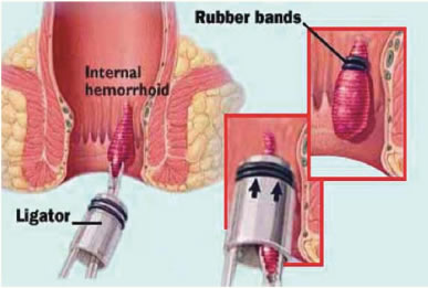
Rubber band ligation
The principle of ligation with rubber bands is to encircle the base of the haemorrhoidal anal cushion with a tight rubber band. The tissue cut off by the rubber band dies and is replaced by an ulcer that heals with scarring. It can be used with first-, second-, and third-degree haemorrhoids. Symptoms can recur several years later but usually can be treated with further ligation.
The most common complication of ligation is pain, but it tends to be mild. However, if the rubber band is applied too distally, the pain is immediate and severe. Bleeding one or two weeks after ligation occurs occasionally and can be severe. Rarely, cellulitis can begin in the tissues surrounding the anal canal. These rare infectious complications may occur in patients who have defective immune systems, from chemotherapy, diabetes to AIDs.
Surgical removal of haemorrhoids (haemorrhoidectomy) usually is reserved for patients with third or fourth-degree haemorrhoids.
During haemorrhoidectomy, the internal haemorrhoids and external haemorrhoids are excised using diathermy. The wounds left by the removal are left open. This is performed as a day case procedure.
Post-surgical pain is the major problem with haemorrhoidectomy. The addition of NSAIDs enhances the relief of pain. Delayed haemorrhage 7 to 14 days after surgery occurs in 1-2% of patients. Wound infections are uncommon after haemorrhoid surgery. Abscess occurs in less than 1% of cases.
However, patients commonly complain of discharge postoperatively and this is expected as the wounds are left open. If the wounds look clean, they do not necessarily need antibiotics. Patients should be advised to keep the area clean and dry and avoid topical applications of ointments or powder.
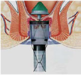
Stapled haemorrhoidectomy is a technique developed in the early 1990s but is a misnomer since the surgery does not remove the haemorrhoids but, rather, the abnormally lax and expanded haemorrhoidal supporting tissue.
The arterial blood vessels that travel within the expanded haemorrhoidal tissue are cut, thereby reducing the blood flow to the haemorrhoidal vessels and reducing the size of the haemorrhoids. During the healing of the cut tissues around the staples, scar tissue forms, and this scar tissue anchors the haemorrhoidal cushions back to their normal position higher in the anal canal — thus, effectively, an anopexy is performed.
However, external haemorrhoids are not removed. Hence, this procedure is best suited for circumferential third or fourth haemorrhoids with minimal external components. It is associated with much less pain than traditional haemorrhoidectomy and patients usually return earlier to work.