Health Resources

Musculoskeletal Oncology

2024/05/16
What is Musculoskeletal Oncology?

Musculoskeletal Oncology includes bone and soft tissue tumours for all age groups, from children to adults. It has three full time musculoskeletal oncologists who have between them more than 50 years experience and 100 publications which have been credited with significant discoveries in musculoskeletal oncology – chief amongst them molecular basis of bone tumour growth and microsurgical joint transfers. It offers a wide range of abilities from prosthetic to bone transplant type options because our specialists hold dual roles as either joint replacement or microsurgical surgeons. It is a unique resource specialised in the care of this very demanding field in South East Asia.

Common Signs & Symptoms

​Presenting history

  • Age
    • Young patients in the first 2 decades of life are prone to primary bone cancers
    • Older patients above 50 years are prone to bone metastasis
  • Pain
    • Unrelenting
    • Disturbs sleep
    • Systemic upset
    • Loss of appetite, loss of weight
    • Shortness of breath
    • Bladder, bowel disturbance

  • Family history
    • Syndromes
    • "General cancer load"
  • Past history
    • Syndromes
    • Cancer history
  • Physical examination
    • General physical examination
    • Springing sign
    • Spine
    • Masses
    • Cutaneous stigmata
    • Café-au-lait
    • Capillary malformations
    • Limb inequality
Common Causes by Age
AgeBenign tumorsMalignant tumorsTumor-like conditions
Birth to 5 yearsEosinophillic granuloma

Leukaemia

Metastatic
neuroblastoma

Osteomyelitis

Non-accidental injury

5 to 15 years

Unicameral bone cyst

Osteochondroma

Aneurysmal bone cyst

Osteoid osteoma

Enchondroma

Non-ossifying fibroma

Chondromyxoid fibroma

Chondroblastoma

Ewing's sarcoma

Fibrous dysplasia

Osteomyelitis

Osteofibrous dysplasia

Stress fracture

15 to 20 years

Unicameral bone cyst

Osteochondroma

Osteoid osteoma

Aneurysmal bone cyst

Nonossifying fibroma

Giant cell tumor

Enchondroma

Chondroblastoma

Chondromyxoid fibroma

Osteogenic sarcoma

Fibrous dysplasia

Diagnosis and Treatment Options
Primary cancers of the bones

Also known as b one sarcomas, osteosarcomas and chondrosarcomas are the two most common variants. These affect individuals of all ages but in particular people who are in their twenties and sixties.

The challenges in managing these conditions revolve around removal of these tumours in a way that they do not recur and then reconstruct the structure to replace the missing anatomical structure and restore function.

The general approaches to reconstruction can be both biological and prosthetic. In biological solutions, the draw is that one is able to reconstruct the missing structure with biological materials and host bone which has a virtually life-long durability. These methods, however, do result in donor site morbidity and have a relatively high early complication rate.

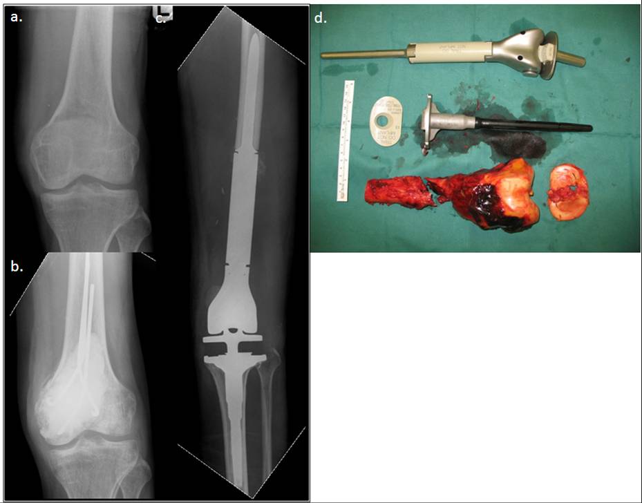
The prosthetic approaches where joints and segments are replaced by metal implants have high patient acceptability and good function but are subject to wear (Figure 1). We have a unique blend of both approaches and hence only the most appropriate is offered in specific cases. Our service has contributed pioneering work in this field with more than thirty years of research in developing novel solutions to the problem. We even have our own in-house bone bank which is the only one of its kind in Singapore and has been responsible for setting up centers regionally.
Primary cancers of the bones
Figure 1. This patient had an osteosarcoma of the lower femur (a). This was biopsied and stabilised and the patient underwent chemotherapy for 9 weeks (b).
The tumour was then resected with oncological sound techniques (ie. margins of resection were clear of tumour) and reconstructed with a tumor megaprosthesis (c). The tumour is replaced with a prosthesis which resembles the lost bone (d). The patient was able to bear weight and walk soon after surgery.

Primary cancers of the soft tissues

Also known as soft tissue sarcomas, liposarcomas and malignant fibrous histiocytomas more commonly occur in adulthood whereas rhabdomyosarcomas and synovial sarcomas occur in childhood.

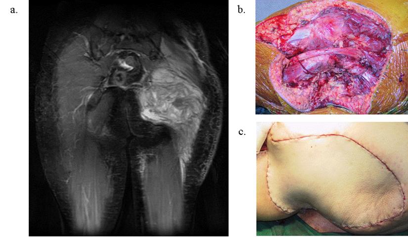
The specific ability of the orthopaedic oncologists is in his ability to resect these tumours primarily to save life and yet secondarily reconstruct defects in a way that maintains function. In addition resections are done in a way that facilitates radiation therapy that is often used in these conditions (Figure 2).
Primary cancers of the soft tissues
Figure 2. This tumour recurred after two previous resections in a non-musculoskeletal oncology specialised unit in an overseas center (a). The patient was seen in our unit for a second opinion. We resected the tumour and using advanced multidisciplinary techniques were able to replace the lost part in a cosmetic fashion (b) without oncological compromise (margins were clear of tumour (c)).

Metastasis

When cancers in other parts of the body spread to the bone and soft tissues, these structures become compromised. As a result patients develop fractures that do not heal and they become invalid. This in turn reduces their life expectancy.

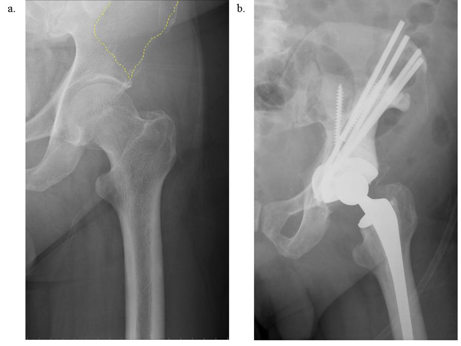
Our specialists have been trained to reconstruct these afflicted bones and joints so that patients with such conditions are able to lead their remaining lives out with dignity (Figure 3). Furthermore, our staff specifically ensures the great sensitivity needed towards such patients.
Metastasis
Figure 3. This patient had cancer of the bladder which had been resected previously. He developed left hip pain and was found to have metastasis to the hip (dotted area in a). Historically, this condition was treated with wheelchair ambulation and radiation. Newer methods which we are able to offer allow such patients to continue ambulation by resecting these tumours and replacing the affected joints (b).

Benign tumours of the bones and soft tissues

These conditions are commonly encountered and treated by general orthopaedists and surgeons. Our special input in this field is in meeting two of the main challenges provided by these conditions.

Firstly, these conditions provide diagnostic problems - that is they can be mistaken for more serious conditions like cancers. Our service works closely with the Departments of Pathology and Radiology with whom we meet regularly and such cases are amply discussed before embarking on surgery. This minimises the risk of mis-diagnoses.

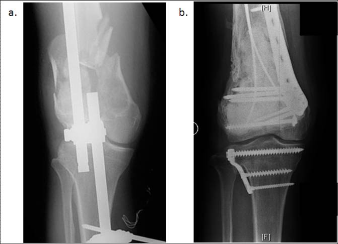
Next, when such conditions compromise function and need to be operated on (eg. Weakening the bone to the point that they may break) we would be able to reconstruct these conditions with minimal functional embarrassment (Figure 4).
Benign tumours of the bones and soft tissues
Figure 4. This patient had a benign condition of the femur which he ignored. Most of such conditions are managed by general orthopaedists at an early stage when they can be effectively controlled. By the time they have broken through the anatomy of the femur is completely disrupted making limb salvage a challenging process (a). We were able to restore the anatomy of the femur by various advanced techniques including the use of bone from our in-house bone bank and the choice of metals in the implants that would not hamper subsequent MRI surveillance for recurrent disease.

Last updated on
Best viewed with Chrome 79.0, Edge 112.0, Firefox 61.0, Safari 11
National University Health System
  • National University Hospital
  • Ng Teng Fong General Hospital
  • Alexandra Hospital
  • Jurong Community Hospital
  • National University Polyclinics
  • Jurong Medical Centre
  • National University Cancer Institute, Singapore
  • National University Heart Centre, Singapore
  • National University Centre for Oral Health, Singapore
  • NUHS Diagnostics
  • NUHS Pharmacy
  • Yong Loo Lin School of Medicine
  • Faculty of Dentistry
  • Saw Swee Hock School of Public Health
Back to Top