Health Resources

Musculoskeletal Trauma Surgery

2024/05/20
Conditions

The Orthopaedic Trauma surgical team provides the following services:

  • Management of acute, severe musculoskeletal trauma, including use of computer-aided surgical planning, minimally invasive fixation (MIPO) and Trauma Navigation for complex fractures and deformities.
  • Reconstruction and joint replacement for periarticular fractures (fractures involving joints).
  • Reconstruction and revision implant surgery for periprosthetic fractures (fractures associated with Total Joint Replacement implants).
  • Management and fixation of complex pelvic and acetabular fractures.
  • Management and fixation of fragility fractures in elderly patients.

Our surgeons also have extensive expertise in the long-term treatment of debilitating post-traumatic sequelae including:

  • Chronic osteomyelitis (infection of the bone);
  • Non-unions (failure of the broken bone to heal);
  • Mal-unions (failure of the bone to heal with appropriate alignment);
  • Joint stiffness following trauma; and
  • Limb length discrepancy (one limb being shorter than the other).

Fractures of the Upper Extremity

  • Shoulder—The joint of the body formed by bones and joints including the soft tissues by which the arm is connected with the trunk.
  • Clavicle—A bone of the (collarbone) pectoral girdle that articulates with the scapula and sternum bones.
  • Scapula—A large triangular (shoulder blade) at the back of the shoulder.

Upper Arm (humerus shaft)
Figure 2. Upper Arm (humerus shaft)

  • The portion of the upper extremity including the humerus (upper arm bones) extending from the shoulder joint to the elbow joint.
  • Humerus—The long bone (upper arm) extending from the shoulder joint to the elbow joint.

Proximal Humerus
Figure 3. Proximal Humerus

  • Elbow—the joint of arm connecting the upper arm bone (humerus) with the forearm bones (radius and ulna).
  • Forearm—the portion of the upper extremity including the radius and ulna (lower arm bones) extending from the elbow joint to the wrist joint.
  • Radius—the bone located on the thumb side of the forearm.
  • Ulna—the bone located on the 'little-finger' side of the forearm.

Factures of the Lower Extremity

  • Hip—the ball-and-socket joint formed by the head of the femur and the cup-shaped cavity of the acetabulum, sometimes used to refer specifically to the proximal femur.

Factures of the Lower Extremity - Hip

  • Femur (upper leg)—the upper bone of the lower extremity that extends from the hip joint to the knee joint.
  • Proximal Femur—the upper portion of the femur.
  • Distal Femur—the lower portion of the femur which forms the upper portion of the knee joint.
  • Factures of the Lower Extremity - Distal Femur

  • Knee—a joint in the middle of the leg that articulates between the femur, tibia/fibula and patella.
  • Patella—a thick triangular bone that is located in the front portion of the knee joint and provides knee stability and joint protection.
  • Tibial Plateau—the upper portion of the tibia which forms the bottom portion of the knee joint.
  • Tibia (lower leg) —the bone located in the inner portion of the lower leg which is the larger of the two bones between the knee and ankle joints.
  • Tibia (lower leg)

  • Proximal Tibia—the upper portion of the tibia.
  • Distal Tibia—the lower portion of the tibia.
  • Pilon—the lower portion of the tibia which (with the distal fibula) forms the upper portion of the ankle joint.
  • Fibula—the bone located in the outer portion of the lower leg which is the smaller of the two bones between the knee and ankle joints.
  • Distal Fibula—the lower portion of the fibula which, together with the tibia, forms the ankle joint.
  • Ankle—the joint between the foot and the lower leg.
  • Calcaneus—the heel bone of the foot.
  • Talus—the bone that bears the weight of the body and forms the lower portion of the ankle joint.
  • Foot—the terminal part of the lower extremity upon which an individual stands.
Other Fractures and Conditions

  • Pelvis—a basin-shaped structure, comprising the pubis in front and the sacrum and coccyx behind, that support the weight of the upper body and transmits weight-bearing to the lower extremities.
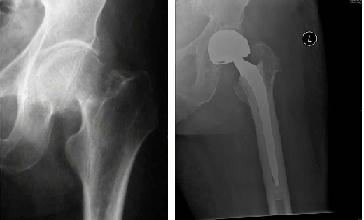
  • Acetabulum—the cup-shaped cavity at the base of the pelvis into which the ball-shaped head of the femur fits (upper portion of the hip joint).
  • Fragility Fractures—occur in osteoporotic bone, generally affecting elderly patients.
  • Polytrauma—a short verbal equivalent used for patients usually with two or more severe injuries in at least two areas of the body.
  • Non-union—failure of a fracture to unite.
  • Mal-union—a bone which heals in a non-anatomic position (out of alignment).
  • Limb Length Discrepancy—describing one limb being shorter than the other.
  • Spine—the series of vertebrae forming the axis of the skeleton that protects the spinal cord.

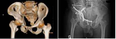
Plevis and Acetabular Fractures

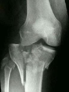
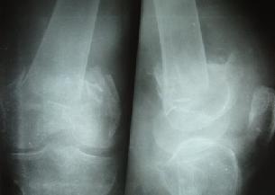
Fractures of the pelvis and acetabulum are among the most serious injuries treated by orthopaedic surgeons. These injuries usually result from high velocity accidents, such as in motor vehicles or high falls. People of all ages are vulnerable to these injuries. Elderly patients with fragile bones due to osteoporosis, in particular, can sustain fractures of the pelvis and acetabulum with a lower impact fall.


Fracture right acetabulum

As with all fractures, we prioritise the patient's ability to return comfortably to normal daily activities. Acetabular fractures, injuring the joint bone and cartilage, occur through the socket of the hip joint. These fractures are often dislocated and require treatment to minimize the fracture. If joints heal with displaced bones, the surfaces will have irregularities, causing excessive wear and resulting in severe joint arthritis, reduced motion and function, along with pain. Proper alignment of the bones during healing is essential. 

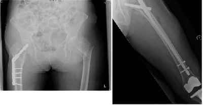
Stable pelvic fracture (no displacement or dislocation) may usually be treated without surgery. Displaced fractures usually require bone. This may be done by either open (surgical) or closed means (non-surgical). Once the bones are realigned with metallic devices including wires, pins, screws, and plates, the surgeon treats the bones internally or externally during healing.

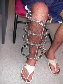
Patients with pelvic fractures may require one or more surgical procedures. After reducing the fracture, the surgeon may use an External Fixation (Ex-Fix) frame to hole the bones in place. Application of an external fixator is done by inserting threated pins into the bone on either side of the fracture. These pins are then connected to rods outside the skin to form a frame.

Ex-Fix technique

While the Ex-Fix technique is sometimes the only procedure needed to repair a fractured pelvis, some patients require additional surgery in which plates and screws are used internally to hold the bones in place. Depending on the site and complexity of the fracture, the surgeon may have to fix the front or back of the pelvis, or both. Separate operations may be needed for each area needing treatment.

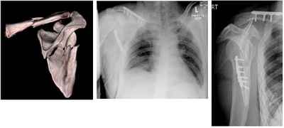
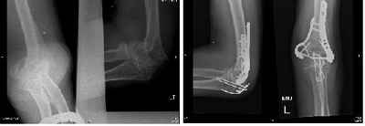
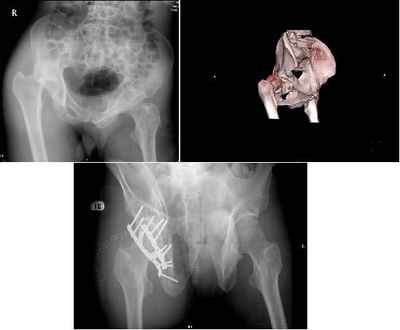
Patients with acetabular fractures often require Open Reduction with Internal Fixation (ORIF), especially those patients with joint displacement. The surgeon realigns the bones precisely to minimize the risk of developing post-injuring related problems, especially arthritis. Plates and screws prevent future displacement and facilitate early rehabilitation.

Treatment for acetabular fractures typically commences 5-10 days after the injury to mitigate the risk of intraoperative bleeding. During this period, patients may be placed in traction as part of an injury prevention program to avoid additional harm.

Complications

 Throughout treatment and recovery, healthcare staff monitor for the following potential complications:
  • Deep vein thrombosis and pulmonary embolism: Blood clots that may form in the veins of the pelvis, thigh and/ or lower legs and may travel to the lungs.
  • Pneumonia: an infection of the lungs that may affect any bed-confined patients who cannot fully expand the lungs as per normal
  • Skin problems resulting from being in one position for an extended period of time
  • Muscle complications due to inactivity.
  • Heterotopic ossification: an abnormal bone formation in an area where there is normally muscle; requiring prompt treatment to prevent this new bone from interfering with joint movement
  • Damaged femoral head: If the joint's articular cartilage lining is affected in a pelvic injury, particularly in acetabulum fractures, the joint surfaces must be kept from rubbing together to reduce the risk of arthritis. Preoperatively, traction or a system of ropes, pulleys and weights are used to relieve pressure in the joint. Surgery with open reduction and internal fixation will securely realign the joints for immediate mobilzzation, preserving the smooth cartilage lining and preventing subsequent arthritis.
  • Avascular necrosis of the femoral head: Patients with dislocated hips or acetabulum fractures, or both, may have disrupted blood flow to the femoral head (the ball in the hip joint). This can result in the death and collapse of bone tissue and hip joint arthritis.
  • Nutritional problems: The body requires more protein and calories during healing.
  • Constipation resulting from inactivity.
  • Infection at the site of the injury.
Fragility Fractures

Osteoporosis-related fractures have become a serious problem, particularly in our region, where there has been a substantial rise in patients requiring treatment for such fractures. Our goal is to achieve secondary fracture prevention in osteoporotic patients through comprehensive investigations, literature review, education, drug treatments and regular monitoring.

Osteoporotic knee fracture in an elderly patient

Minimally Invasive Procedures

Our musculoskeletal trauma surgeons approach each patient case as conservatively as possible. However, when surgical treatment is required, we advise the most minimally invasive solution for the speediest recovery.

Some of the techniques we employ when treating various fractures include the following:

  • Intramedullary Nailing: Nails / Rods are inserted into the bone marrow canal in the center of long bones of the extremities (commonly the femur or tibia) to align and stabilize the limb. These rods share the weight with the bone, an advantage that allows patients to recover more quickly.
  • Plate and Screws: Surgeons fix the bone fragments to their proper positions to stabilize the bone until it heals. Previously, using plates and screws to fix fractures required extensive incisions. Now, our surgeons are trained to perform minimally invasive plate fixing through small incisions.
  • External Fixation: This method is often chosen for bones with infections or loss of bone mass due to an open fracture (compound) and used to immobilise the bone for proper healing. Rigid support with pins or screws placed into the bone on both sides of the fracture achieve that purpose. An external apparatus bridging across the broken area may also be used to correct the shortening or angulation of an extremity.
Correction of Deformity and Limb Length Discrepancy

Individuals with shortened extremities or abnormal extremity alignment from a prior injury can opt for corrective surgery.

Limb lengthening and reconstruction techniques, utilising intramedullary nails and possibly plates and external fixing, are applied to replace missing bone and to lengthen or straighten deformed bone segments of legs and arms. Deformities, secondary to osteoarthritis, can also be corrected to achieve pain relief and delay arthritis.


Correction of deformity
A circular external fixator applied for correction of deformity

Doctors
Last updated on
Best viewed with Chrome 79.0, Edge 112.0, Firefox 61.0, Safari 11
National University Health System
  • National University Hospital
  • Ng Teng Fong General Hospital
  • Alexandra Hospital
  • Jurong Community Hospital
  • National University Polyclinics
  • Jurong Medical Centre
  • National University Cancer Institute, Singapore
  • National University Heart Centre, Singapore
  • National University Centre for Oral Health, Singapore
  • NUHS Diagnostics
  • NUHS Pharmacy
  • Yong Loo Lin School of Medicine
  • Faculty of Dentistry
  • Saw Swee Hock School of Public Health
Back to Top