Deep tech innovations like Mammosense and FxMammo, co-developed by NUH,
are making breast cancer screening more accurate and comfortable,
which in turn facilitates earlier detection and timely treatment.
Issue 7 | December 2024

 Subscribe and ensure you don't miss the next issue!
Breast cancer is the most common cancer among women in Singapore, making up approximately 30 per cent of all cancer diagnoses. Each year, around 2,000 women are diagnosed, with about 400 lives lost to the condition.
Early detection is key to beating breast cancer, with mammography remaining as the gold standard screening technique. Nevertheless, despite recommendations from the Health Promotion Board, screening rates in Singapore have remained low at about 40 per cent. Factors such as physical discomfort and the lengthy diagnostic process deter many women from regular screenings, often leading to delayed diagnoses.
The National University Hospital (NUH) has co-developed deep tech innovations, including the LiDAR-enabled Mammosense and artificial intelligence (AI)-powered FxMammo, designed to ease the concerns and anxieties surrounding breast cancer screening.

Reducing physical discomfort
Traditional breast cancer screening can be physically uncomfortable, often discouraging women from undergoing the procedure.
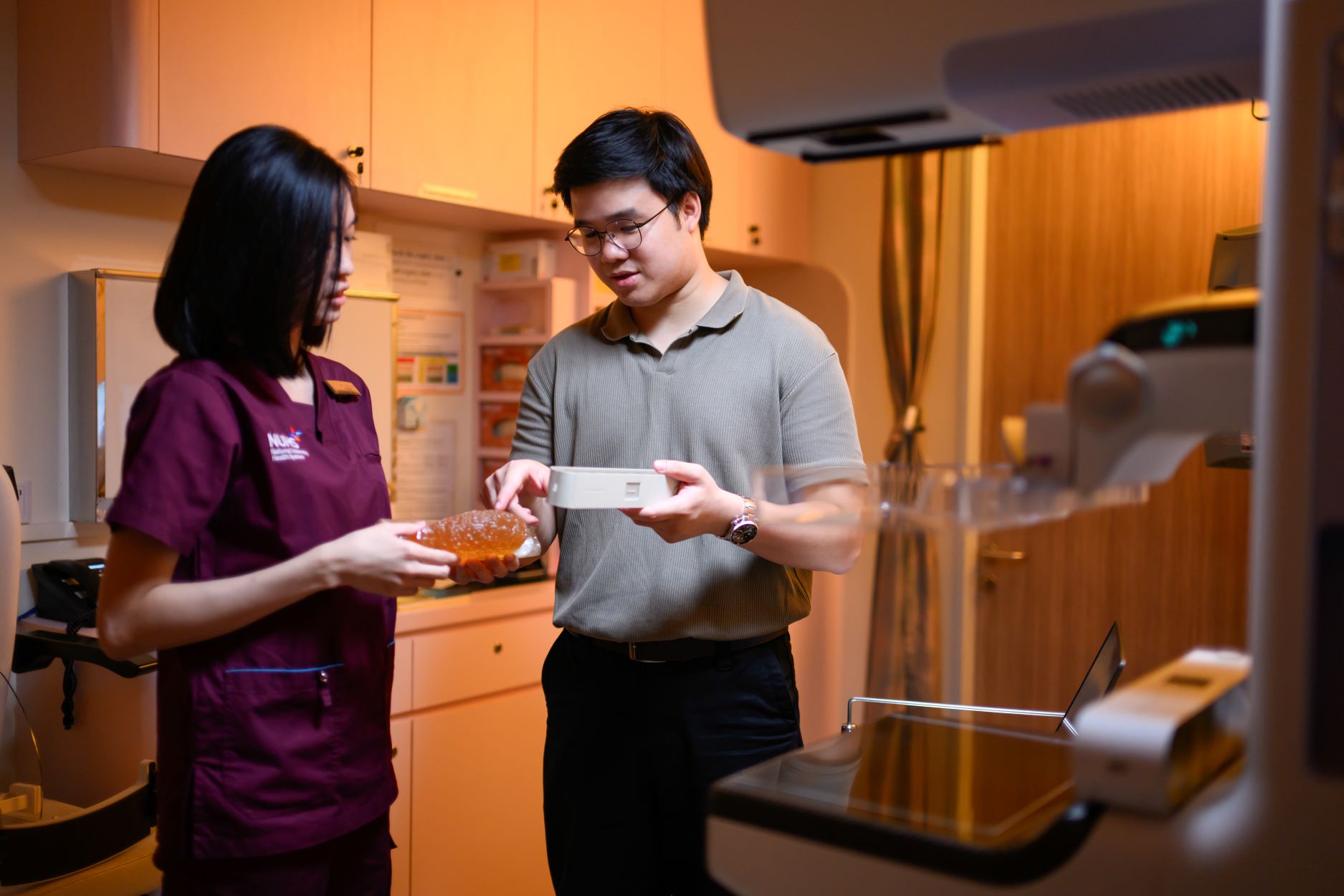
In current practice, radiographers ask patients to remain still as a compression plate flattens each breast against an X-ray machine. This process is repeated several times as each breast is repositioned to capture images from various angles. The radiographers rely on their experience to estimate the compression force needed on each breast, which can lead to over- or under-compression.
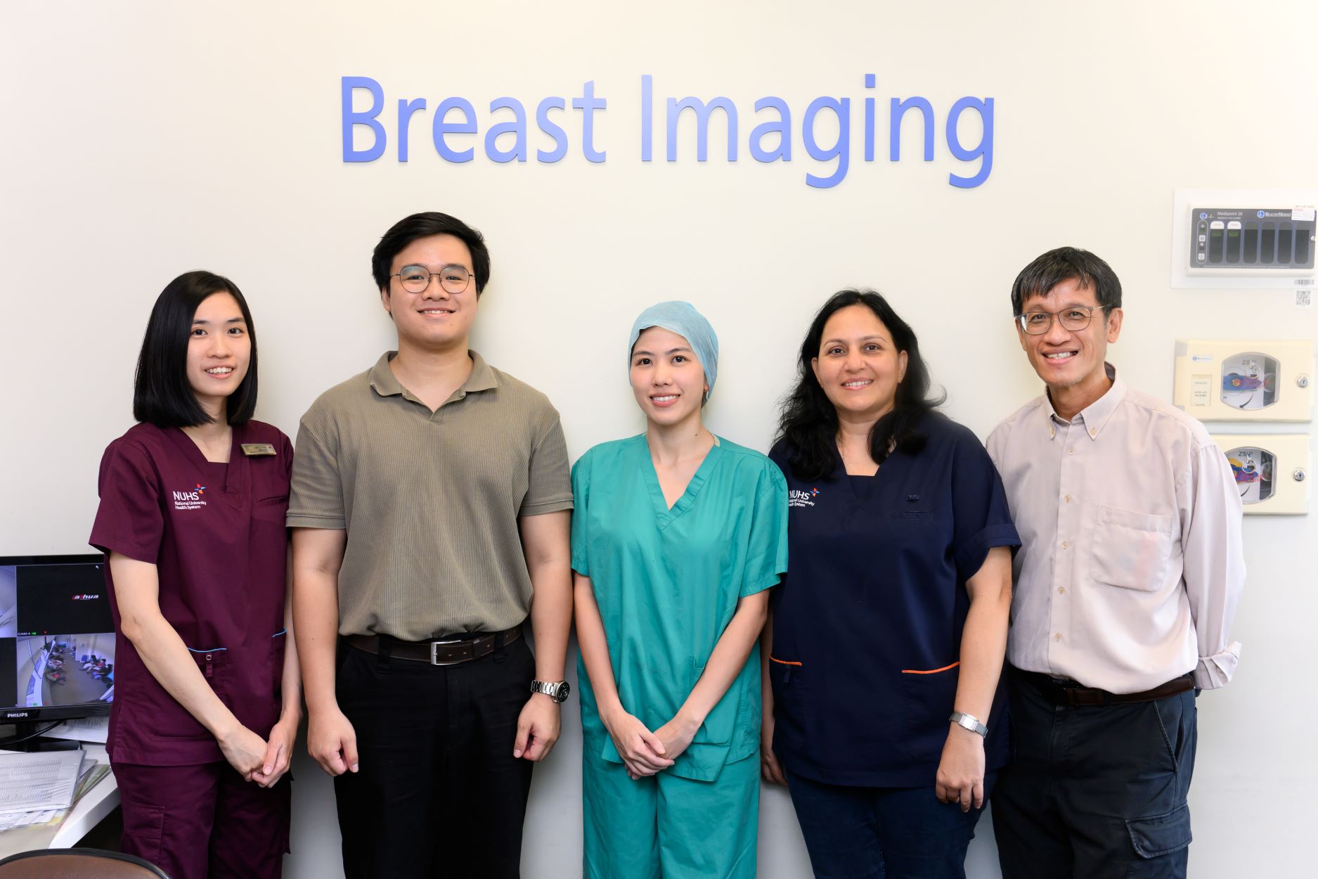
Mammosense was developed as a tool to guide radiographers conducting personalised breast compressions, and thus reduce a woman’s discomfort during screenings. The tool, co-patented by NUH together with Mr Luke Goh and the National University of Singapore, was named the Singapore winner for the 2024 James Dyson Award. The NUH development team included Dr Pooja Jagmohan, Senior Consultant, and Ms Lim Li Ying, Senior Radiographer from the Department of Diagnostic Imaging, NUH, as well as Dr Serene Goh, Associate Consultant, Department of Surgery, NUH.

Powered by a light detection and ranging system called LiDAR, Mammosense allows radiographers to get a customised reading of how much compression force they should use on each person. Early trials show the technology reduces force exertion by 34 per cent and pain by 25 per cent.
“This enhances comfort and encourages more women to undergo screenings. On the other hand, the technology also gives radiographers greater confidence in their work without the constant worry of causing unnecessary discomfort to patients,” says Ms Lim.
Streamlining the detection process
Apart from physical discomfort, conventional screening methods generate mammograms in which both dense breast tissues and potential anomalies register as similar shades of white, obscuring tell-tale signs of breast cancer. This can complicate diagnosis, sometimes delaying reports for weeks. Mammograms also risk missing 20 per cent of breast cancers present at the time of screening.
Associate Professor Mikael Hartman, Head & Senior Consultant, Division of General Surgery (Breast Surgery) Department of Surgery, National University Hospital, co-developed FxMammo to enhance the accuracy and efficiency of breast cancer screening through advanced image analysis.
“FxMammo effectively and reliably aids radiologists in detecting breast cancer on digital mammograms. The AI model in FxMammo analyses images and provides radiologists with a heat map and a cancer probability score. This map helps radiologists pinpoint the areas FxMammo used to calculate its score,” says A/Prof Hartman.
Now being trialled in more than 30 hospitals and universities across Asia, FxMammo is as accurate as a trained radiologist. “Using a metric called ‘area under the curve’, where 1.0 is perfect, a radiologist scores about 0.9, while FxMammo’s score is between 0.91 and 0.93,” A/Prof Hartman adds. “With half of the world’s population being women who need mammograms — plus a shortage of radiologists globally — the need for such a technology is huge.”
Pushing the frontiers of breast cancer research
Breast cancer teams at NUH continue to push the frontiers of AI-integrated breast cancer research.
One recent study explored the perspective of radiologists on incorporating AI into Singapore’s breast screening programme to identify key barriers and facilitators. The findings highlight the importance of comprehensive training, legal safeguards and collaboration between AI developers, healthcare providers and policymakers.
Another study zoomed into AI governance in breast cancer screening, which provided valuable insights into the essential components required to establish a safe and effective framework for AI adoption.
In a separate study using FxMammo, researchers demonstrated that the AI-powered software improved the diagnostic accuracy of resident radiologists, bringing their performance closer to that of consultants. Beyond bridging gaps in expertise, the software could potentially save an approximately SGD 2.7 million each year due to the time savings achieved in diagnosing non-malignant cases.
Like this article? Simply subscribe to make sure you don't miss the next issue of EnvisioningHealth!


