- Bone Mass Densitometry
- Breast Imaging
- Cardiac Imaging
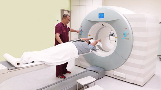
- Computed Tomography
- Fluoroscopy
- Intravenous Urography
- Interventional Radiology
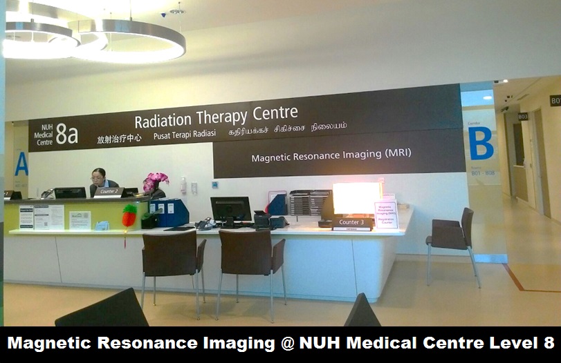
- Magnetic Resonance Imaging
- Nuclear Cardiology
- Nuclear Medicine
- PET/CT
- Plain X-ray
- Ultrasound
- Vascular Imaging
Breast Imaging
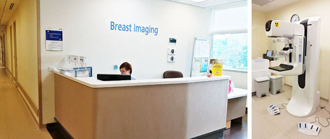
Breast Imaging serves as the primary method for early detection and diagnosis for breast diseases. Our department’s Breast Imaging Centre is dedicated to providing the following services:
- Assessment centre for Breast Screen Singapore (BSS)
- Screening and diagnostic mammography
- Ultrasound breast
- Ultrasound axilla
- Ultrasound-guided breast biopsy
- Mammogram-guided breast biopsy
- Image-guided fine needle aspiration
- Image-guided hookwire localisation
Computed Tomography (CT) is an advanced X-ray technique that combines computer technology with X-rays to generate high-resolution 3D images of the internal organs. These detailed 3D images enable radiologists and clinicians to make more precise diagnosis and plan surgical interventions effectively.
Fluoroscopy involves the use of X-rays to capture real-time, cine-like images of an organ while it is functioning. This is achieved through a specialised X-ray machine consisting of an exploratory X-ray unit and a corresponding image intensifying plate. Fluoroscopy is applicable to imaging various parts of the body and is particularly useful in gastrointestinal imaging.
Some common fluoroscopic procedures include:
- Barium meal and follow-through
- Barium enema
- Micturating cystogram (MCU)
- Hysterosalpingogram (HSG)
Intravenous Urography , commonly known as IVU or IVP, is an X-ray examination of the urinary system where a contrast media ( X-ray dye) is used. It can aid in diagnosing obstructions or abnormalities in the kidneys, ureters and bladder.
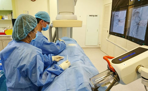
Interventional Radiology is a subspecialty of radiology that involves the performance of minimally invasive procedures guided by imaging. Some of these procedures serve diagnostic purposes (e.g., angiogram), while others are therapeutic (e.g., angioplasty).
During these procedures, imaging is used to direct the use of needles or other tiny instruments, such as catheters. These images act as road maps, enabling the Interventional Radiologist to navigate the instruments through the body to reach specific areas of interest.
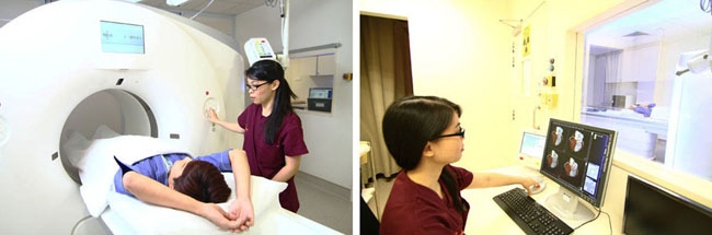
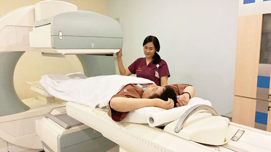
Magnetic Resonance Imaging
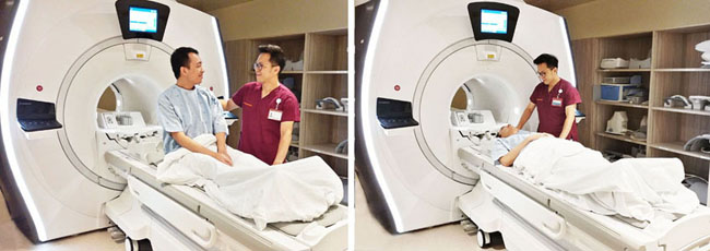
Magnetic Resonance Imaging (MRI) is a diagnostic technique that uses powerf ul magnetic field and radio waves instead of X-rays. This technique provides clear and detailed images of internal organs and tissues.
Some common MRI scans include:
- MRI Brain/Head/Neck
- MRI Spine/ Musculoskeletal
- MRI Vascular
- MRI Body
Nuclear Medicine is a specialised field in medical imaging that diagnoses the selective uptake of radiopharmaceuticals by various bodily systems. This modality is sensitive to physiological and metabolic changes, making it a key tool in the diagnosis of various types of cancers, heart diseases, endocrine disorders and other abnormalities.
PET/CT integrates two imaging modalities by combining functional information from a Positron Emission Tomography (PET) with anatomical information from Computed Tomography (CT) into a single study. A PET examination can detect changes in cellular function, revealing how your cells utilise nutrients such as sugar and oxygen. Since functional changes occur before physical changes, information from PET aids doctors in early diagnosis. The integration of PET and CT allows more precise information for diagnosing the presence and extent of disease, as well as prescription of treatment and therapy progress monitoring.
Clinical applications:
- Cancer
- Inflammation
- Other malignancies
- Other diseases
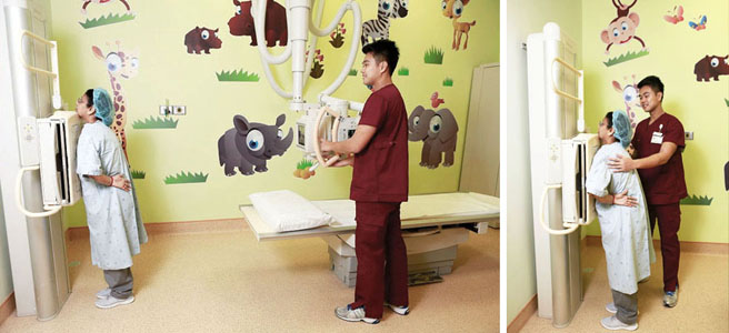
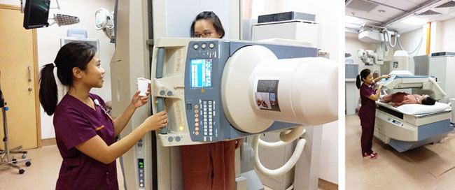
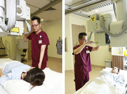
Plain X-ray is used to reveal the internal structure of the body, with the amount and strength of radiation varying based on the specific body part being imaged.
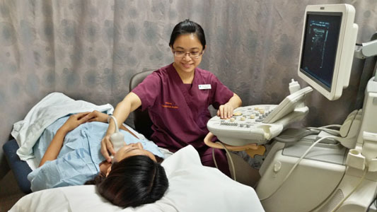
Ultrasound involves the use of high-frequency sound waves to generate images of internal organs. Some common ultrasound scans include:
- General Ultrasound
- Vascular Ultrasound
- Transplant Related Study
- Paediatric Ultrasound Study
- Musculoskeletal Ultrasound Study

 Diagnostic Imaging @ KRW Level 3
.jpg?sfvrsn=970ea959_1)