NUH offers a range of routine diagnostic and therapeutic procedures for managing digestive and liver diseases. Among these is the Helicobacter Pylori Infection – Carbon (C13) Urea Breath Test, along with various other procedures available at our centre.
What is a Gastroscopy?
Oesophago-Gastro-Duodenoscopy (OGD), commonly known as Gastroscopy, is a procedure enabling doctors to examine the lining of the oesophagus, stomach and the duodenum (the initial segment of the small intestine). This examination is performed in the endoscopy suite using a soft, flexible tube, which is thinner than a finger, gently inserted through the mouth. The procedure employs a video camera, allowing the doctor to thoroughly inspect the upper gastrointestinal tract.
Who should have a Gastroscopy?
Gastroscopy is advisable for individuals needing:
How do I prepare for one?
Patients must fast for at least six hours before the procedure to ensure an empty stomach, crucial for effective gastroscopy. An empty stomach provides clear visualisation and reduces vomiting risks during the examination. The presence of food may necessitate rescheduling the procedure.
Patients should consult their doctor regarding medication continuation, particularly for blood thinners, warfarin, aspirin or diabetes medication.
How is a Gastroscopy performed?
The procedure begins with the administration of a local anaesthetic spray to numb the throat, followed by optional light sedation for relaxation. The doctor then gently advances the gastroscope though the mouth into the stomach. The entire process typically takes about 10 minutes.
Patients might experience mild bloating or belching due to air inflation in the stomach. The procedure is pain-free, well-tolerated and does not affect breathing.
What happens after a Gastroscopy?
Post-gastroscopy, patients are observed until the effects of sedation subside and are then discharged. It is advised not to drive or operate machinery until the next day due to sedative effects.
Mild bloating from the introduced air and occasional mild sore throat may occur, usually resolving within a few days. Normal eating can resume on the same day and regular activities can continue the following day.
What are the risks?
Gastroscopy is a safe procedure with infrequent complications. However, risks include bleeding from a biopsy site, perforation or aspiration of stomach contents. Fasting minimises aspiration risks. Some patients may react to sedation. In cases of severe chest or abdominal pain, fever, or bleeding, it is crucial to contact a doctor immediately.
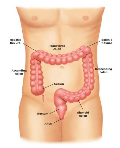
Who should have a Colonoscopy?
Colonoscopy is recommended for individuals needing:
How do I prepare for one?
For an effective colonoscopy, the colon must be completely empty. This generally involves bowel cleansing, for which instructions will be provided. The preparation may include consuming polyethylene glycol or laxatives, followed by water to cleanse the bowel. Incomplete preparation may necessitate rescheduling the procedure.
Patients should discuss medication continuation with their doctor, particularly for blood thinners warfarin, aspirin or diabetes medication.
How is a Colonoscopy performed?
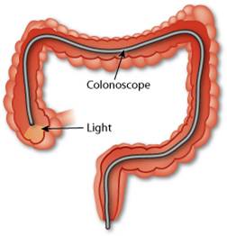
Light sedation is administered for relaxation and to alleviate discomfort. Patients may feel pressure, bloating or cramping during the procedure, but these sensations are generally well-tolerated. The doctor will carefully advance a colonoscope though the colon to inspect its lining. The procedure typically lasts 15 to 30 minutes.
What happens after a Colonoscopy?
After the colonoscopy, patients are monitored until sedation effects subside, followed by discharge. Operating machinery or driving is not advised until the next day due to the sedative effects, which can impair reflexes.
Mild cramping or bloating from the air introduced into the colon may occur, improving rapidly once gas is passed. Normal eating can resume the same day, and regular activities can continue the following day.
What are the risks?
Colonoscopy is a safe procedure with rare complications. These may include bleeding (less than 1%) and bowel perforation (0.2%). Bleeding can occur up to two weeks post-procedure, and in rare cases, surgical intervention may be necessary. Some patients might react to sedation. In the event of severe abdominal pain, fever, or rectal bleeding post-colonoscopy, it is imperative to contact a doctor immediately.
What is EUS?
EUS enables doctors to examine the walls of the upper and lower gastro-intestinal tract, including the oesophagus, stomach, duodenum, colon and rectum. EUS is also used to study adjacent internal organs, such as the bile duct and pancreas. The procedure involves using a slender, flexible endoscope, inserted through the mouth or anus to the examination site. An ultrasound component then generates sound waves to create detailed images of the digestive tract.
Why is EUS done?
EUS provides detailed pictures of the digestive tract, aiding in diagnosing conditions causing abdominal pain or unexplained weight loss. It can confirm diagnoses where other conditions have been ruled out, providing additional reassurance.
EUS is also essential for evaluating abnormalities, such as growths identified in previous endoscopies or X-rays, giving detailed insights into their nature and assisting in treatment planning. In addition, EUS can be used to diagnose diseases of the pancreas, bile duct and gallbladder when other tests are inconclusive.
How should I prepare for EUS?
Upper tract EUS: Fasting, including abstaining from water, is usually required six hours before the examination. Specific fasting instructions will be provided.
Lower tract EUS: This may involve consuming a special cleansing solution or following a clear liquid diet with laxatives or enemas. Failure to adhere to preparation instructions may lead to rescheduling.
Discuss medication continuation with your doctor, especially for blood thinners like warfarin, aspirin, ticlopidine, clopidogrel and medication for high blood pressure or diabetes.
How is EUS performed?
The experience of undergoing EUS is similar to OGD or colonoscopy, depending on whether the upper or lower gastrointestinal tract is being examined.
What happens after EUS?
Post-EUS, patients are monitored until sedation wears off before being discharged. Avoid driving or operating machinery until the next day due to sedative effects, which may impair reflexes. A mild sore throat may occur, resolving within a few days. Normal eating and activity resumption is typically possible on the same day.
What are the risks?
While complications are rare, they can include bleeding from a biopsy site, perforation, aspiration of stomach contents, reactions to sedatives, infections and complications related to heart or lung diseases. The risk slightly increases if deep needle aspiration is performed during EUS. It's crucial to weigh these risks against the procedure's benefits and the risks of alternative approaches.
In case of severe chest or abdominal pain, fever or bleeding post-EUS, consult your doctor immediately.
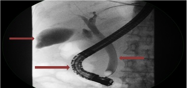
ERCP is a specialised endoscopic technique used to diagnose and treat conditions of the biliary tree, which includes the gallbladder, bile ducts, and pancreatic ducts. ERCP combines endoscopy and X-ray imaging, enabling physicians to acquire detailed images of these structures. The procedure involves inserting an endoscope through the mouth to access the small intestine, where the openings of the bile and pancreatic ducts are located.
ERCP is often employed to identify strictures (narrowing) in the bile and pancreatic ducts, perform biopsies of abnormalities and remove gallstones. During the procedure, a plastic or metal stent may be placed within the bile or pancreatic ducts to facilitate drainage.
What is a liver biopsy?
A liver biopsy involves removing a small sample of liver tissue for microscopic examination. This is typically done using a needle inserted into the liver to extract a tissue sample for analysis.
Why do I need to have a liver biopsy?
A liver biopsy assists doctors in diagnosing specific liver conditions and determining their severity, which is vital for guiding treatment. Your doctor will discuss in detail the reasons for the biopsy prior to the procedure.
Are there complications to doing a liver biopsy?
While liver biopsies are generally safe, some complications may occur. These include:
These complications are rare and typically managed without surgery. However, in very rare cases, complications from a liver biopsy can lead to surgery or even death.
How is a liver biopsy performed?
Liver biopsies can be performed either bedside by a liver specialist or under ultrasound guidance by a radiologist. Typically, this outpatient procedure takes place in the day surgery ward.
During the biopsy, you will lie on your back while a specific skin area is targeted with ultrasound assistance. After cleaning the area, a local anaesthetic is applied to numb the skin and underlying tissue. The doctor will guide your breathing, instructing when to hold your breath. At this moment, a thin needle is inserted through the skin into the liver, quickly extracting a tissue sample. The process takes only one to two seconds. To ensure comfort, medication may be administered. The extracted tissue is then sent for laboratory analysis. The entire procedure lasts approximately 15 to 20 minutes.
What will I expect on the day?
On the day of the biopsy, you will be admitted to the day surgery ward in the morning. Blood tests may be conducted to ensure the safety of the procedure. After undergoing the liver biopsy, as described, you will be monitored in the ward for four to six hours. During this period, nurses will check your blood pressure, heart rate and manage any pain with painkillers. A doctor will review and examine you before discharge. The total duration from admission to discharge can be up to eight hours.
Will there be serious damage to my liver?
As the biopsy involves removing only a small piece of liver tissue, it does not cause significant damage to the liver.
What do I need to watch out for?
Post-procedure, patients should avoid strenuous physical activity and lifting heavy weights for two weeks. A medical certificate for rest will be issued. If you experience faintness, blackouts, palpitations or severe pain after returning home, seek immediate medical attention.
Are there any other alternative approaches to a liver biopsy?
An alternative method is the transjugular liver biopsy, involving a needle passed through a tube in a neck vein, extracting liver tissue through the blood vessel. This method poses an even lower risk of bleeding and is safer for patients with abnormal blood clotting. However, it may yield less diagnostic tissue and is more costly. This approach may be recommended for patients with a high risk of bleeding.
For any questions about liver biopsy, please contact one of our hepatologists.
For information on other specialist endoscopic procedures and motility and functional investigations, please click here.